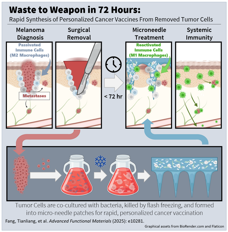
We've published breakthrough research in Advanced Functional Materials that could transform how we prevent cancer from coming back after surgery. Our team developed a personalized cancer vaccine that can be produced in just 72 hours using the patient's own tumor cells—a dramatic improvement over current cell therapies that take 6-8 weeks.
Here's how it works: we take tumor cells removed during surgery and engineer them with an attenuated bacteria strain to increase the response they trigger when exposed to immune cells. These modified cells are then combined with L-arginine and delivered through dissolving micro-needle patches that patients can receive right at the surgical site. The system works through three mechanisms: it amplifies presentation of the patient's specific tumor antigens, reprograms immune-suppressive macrophages into tumor-fighting cells, and generates nitric oxide that triggers inflammatory cell death in any remaining cancer cells. Specifically, the bacterial components activate the NF-κB signaling pathway, which drives macrophages to produce high levels of iNOS—an enzyme that converts the supplied L-arginine into toxic nitric oxide that kills tumor cells. When we tested this approach in mouse models of incomplete melanoma resection, the results were remarkable. The vaccine reduced tumor regrowth by 92.7% without any other treatment, including complete remission in 20% of cases. Even more importantly, we show these vaccines have a strong abscopal effect meaning that the immune system learns to fight cancer cells anywhere in the body, not just at the micro-needle application site. What makes this especially promising is the speed and personalization. Unlike generic cancer vaccines, this approach uses each patient's actual tumor cells, ensuring coverage of their unique cancer signature. The rapid 72-hour production timeline means treatment can begin while the immune system is still primed from surgery. We are excited about expanding this work out to other cancer types in the future.
0 Comments
I'm excited to share that our team's latest paper on statistical image processing for cancer margin detection during surgery has been accepted in Analytical Chemistry.
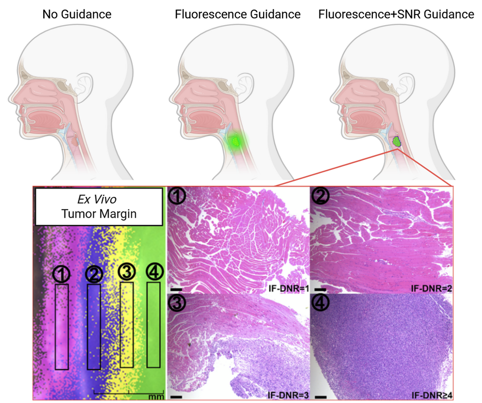
Our work reveals a strong correlation between tumor and surrounding tissue fluorescence variance, enabling stable tumor boundary identification for up to 24 hours post-injection - a key finding that could help to standardize the window for guided resections. By stratifying fluorescence signal by standard deviations above background, we demonstrated that SNR thresholds correlate strongly with cancer probability. The approach was validated first in xenograft models and then successfully translated to clinical samples through our collaboration with China Medical University, achieving millimeter scale diagnosis with 87.5% accuracy with no false negatives. This method offers potential for precise, real-time, quantitative guidance during cancer resection to help surgeons ensure complete tumor removal while preserving healthy tissue. I'm excited to share our latest research published in Cell Reports Physical Science on a game-changing advancement in photothermal therapy for cancer treatment. Our team discovered that the dimer of indocyanine green (ICG), ICG-II, can completely eliminate tumors in just 2 minutes, compared to regular ICG which doesn't work even after 5 minutes.
The key breakthrough centers around what we call "butterfly oscillation." ICG-II is essentially two ICG molecules linked together, and this pairing creates a constrained molecular motion that dramatically improves how efficiently light energy converts to heat. While regular ICG only converts 47% of laser energy to heat, ICG-II achieves an incredible 95.6% conversion efficiency - the highest ever reported for a small-molecule photothermal agent. Here's what makes this so exciting: the molecular structure of ICG-II forces it to undergo a "butterfly" vibration where parts of the molecule flap toward and away from each other. This motion facilitates non-radiative energy release as heat rather than fluorescence, making it vastly more effective at destroying cancer cells through targeted heating. In our mouse studies, direct injection of ICG-II followed by just 2 minutes of near-infrared light completely eliminated xenograft tumors in some cases, with tumor temperatures reaching over 60°C. The treatment was so effective that two mice experienced complete remission. Meanwhile, equivalent doses of regular ICG couldn't achieve tumor clearance even with longer treatment times. What's particularly promising is that ICG-II maintains excellent biocompatibility and is actually a known, FDA-permitted compound that forms naturally as an impurity during ICG production. This could significantly accelerate the path toward clinical applications, potentially offering cancer patients a faster, more effective photothermal treatment option. We've developed a promising new tool that could make oral cancer surgery more precise. Our team created a specialized fluorescent dye called LP-S that lights up metastatic lymph nodes, making them easier for surgeons to identify and remove.
Right now, surgeons rely on a standard dye called ICG, but it's not very specific - it highlights everything, making it hard to tell which lymph nodes actually contain cancer cells. Our new dye is much more precise, specifically binding to EGFR receptors, which are found in high concentrations on oral cancer cells. When we tested LP-S in mouse models, the results were impressive. The dye produced clearer, longer-lasting fluorescence in cancerous lymph nodes compared to ICG, giving surgeons a much better view of what they're working with. This means they can more accurately remove the correct lymph nodes while leaving healthy tissue in place. If this technology makes it through clinical testing, it could be a game-changer for oral cancer patients. Better lymph node mapping means more accurate cancer staging and ultimately better treatment outcomes. We've been working with our colleagues at IceKredit on an exciting project for the First Global AI Drug Design Competition sponsored by Baidu and Tsinghua University to help identify potential COVID-19 treatments. Using advanced machine learning, we developed a model that can predict whether new compounds might be effective against SARS-CoV-2.
What makes our approach special is that it looks at molecules from multiple angles - both the fine details of their structure and their broader chemical fingerprints. This multi-scale view helps us make more accurate predictions about which compounds are worth testing in the lab. This work builds on our recent publication in the Journal of Computer-Aided Molecular Design and shows how our computational methods can be applied to tackle urgent health challenges like the pandemic. Bayesian regression model combining docking and deep generative networks published in JCAMD8/8/2023 We've published new research in the Journal of Computer-Aided Molecular Design that could speed up the hunt for new medicines. Our team developed a hybrid AI model that's better at designing potential drug molecules than traditional computational methods.
Here's how it works: we combined two powerful AI approaches - one that generates new molecular structures and another that predicts how well those molecules will bind to their target protein. By linking these together with reinforcement learning, our system can efficiently explore vast chemical possibilities and focus on the most promising candidates. When we tested this approach on DDR1 kinase (a protein involved in cancer), the results were impressive. Our hybrid model found molecules with much higher binding scores than conventional similarity-based methods. Even better, it discovered chemically diverse compounds that looked quite different from known inhibitors but still bound effectively to the target. What makes this especially exciting is that our approach reduces one of the biggest limitations in AI drug discovery - the need for massive datasets. Instead of just making slight tweaks to existing drugs, our system can venture into unexplored chemical territory to find genuinely novel compounds. While there's still more work ahead, this hybrid strategy could help researchers identify promising new treatments for diseases where current drugs aren't working well enough. I'm excited to share that our collaborative work with Professor Hui Wei's team at NJU-BME has been recognized with the RSC Horizons Dalton Prize. This interdisciplinary project focused on advancing our understanding of nanozymes and their potential applications.
Nanozymes are artificial nanomaterials that can act like natural enzymes - they catalyze reactions just like the biological enzymes in our bodies, but they're engineered rather than evolved. The potential applications are pretty exciting: these synthetic catalysts could be used to treat cancer and inflammation, and they're even being explored for wearable medical devices. Our research demonstrated that these designed nanozymes actually outperform natural enzymes in several key areas. The work also provided new insights into what factors control how effectively these artificial catalysts work - knowledge that will be crucial for designing even better nanozymes in the future. This recognition highlights both the innovative science and the successful international collaboration that made this research possible. It's a great example of how interdisciplinary partnerships can lead to breakthroughs with real-world biomedical applications. |
Author
Chris Archives
June 2025
Categories |